 Platelets are small bits of cells that aid in the formation of blood clots and help seal breaks in blood vessels.
They are formed by pinching off small bits of a large cell called a megakaryocyte. Megakaryocytes are found in bone marrow. The platelets contains mitochondria and cytoplasm but no nuclei.
Platelets have a number of enzymes and factors that promote wound healing and their membranes are studded with various receptors and factors that promote blood clotting at the site of injury. Normal blood has a very high concentration of platelets (200,000 per microliter) - this is the platelet count that's a common diagnostic test for many medical problems. Platelets have a half life of about ten days so they need to be continuously produced in the bone marrow.
 When a blood vessel is injured a patch of endothelial cells are destroyed exposing the underlying collagen matrix. Platelets bind to collagen and then to each other leading to an aggregation of platelets and formation of a plug that stops the bleeding. The platelet plug also stimulates blood clotting at the site of injury because many of the factors that promote clotting are carried by platelets.
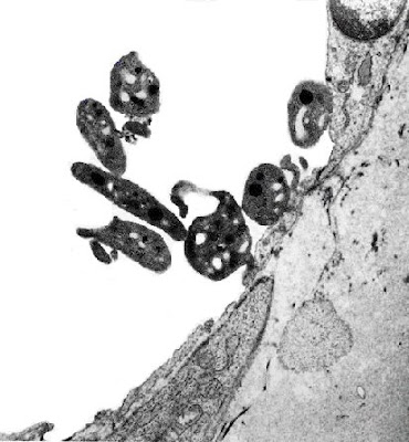
This process is shown in the electron micrograph on the right. The platelets are the small dark cell-like objects. Some of them have adhered to the collagen matrix on the far right and this stimulates other platelets to bind to the ones that first arrived at the lesion. A platelet plug is building.
These platelets will also become activated for formation of fibrin blood clots at this site. Many of the proteins on the cell surface will aid in generating thrombin from prothrombin. Thrombin cleaves fibrinogin to produce the clotting protein, fibrin [Blood Clotting: The Basics].
Gary Carlson has created a number of very impressive images of platelets. Click on the images of aggregating platelets and blot clots forming at a wound.
(Electron micrograph is from Platelets)
No comments:
Post a Comment